Home
/ Posterior Pelvis Anatomy Muscles / Pelvis And Hip Joint Amboss : The pubococcygeus is the intermediate part of the levator ani muscles.
Posterior Pelvis Anatomy Muscles / Pelvis And Hip Joint Amboss : The pubococcygeus is the intermediate part of the levator ani muscles.
Posterior Pelvis Anatomy Muscles / Pelvis And Hip Joint Amboss : The pubococcygeus is the intermediate part of the levator ani muscles.. Tibialis posterior muscle (musculus tibialis posterior) tibialis posterior is the most central and deepest muscle located in the posterior aspect of the leg. Whenever someone talks about the pelvic floor in general, they are probably talking about these 5 muscles: The pelvic floor is also known as the pelvic diaphragm. These muscles originate near the anteroinferior external surface of the bony pelvis and insert at the linea aspera. Lying exposed between the protective bones of the superiorly located ribs and the inferiorly located pelvic girdle, the muscles of this region play a critical role in protecting the.
3 illustrations of the anterior thigh region detail the anatomy of the femoral quadriceps muscle (rectus femoris, vastus medialis, vastus lateralis and vastus intermedius muscles), the gracilis, sartorius and. The muscles of the abdomen, lower back, and pelvis are separated from those of the chest by the muscular wall of the diaphragm, the critical breathing muscle. The pelvic cavity and perineum. The levator ani muscles are the largest group of muscles in the pelvis. Its primary role is in arm movement,.
Anatomy Of The Pelvis Prof Saeed Abuel Makarem from slidetodoc.com It supports the retroperitoneal viscera and neurovascular structures. There are many muscles that form the pelvic floor, including puborectalis, pubococcygeus, iliococcygeus and coccygeus. Biceps femoris long and short head, semitendinosus, and semimembranosus. The gastrocnemius is the most superficial of the muscles and has two heads, medial and lateral. These three muscles are collectively referred to as the hamstring muscles. They have several functions, including helping to support the pelvic organs. Together with popliteus, flexor hallucis longus and flexor digitorum longus, it forms the deep group of muscles of the posterior compartment of leg. The gluteus maximus attaches from the posterior iliac crest, posterolateral sacrum, and coccyx to the gluteal tuberosity and itb.
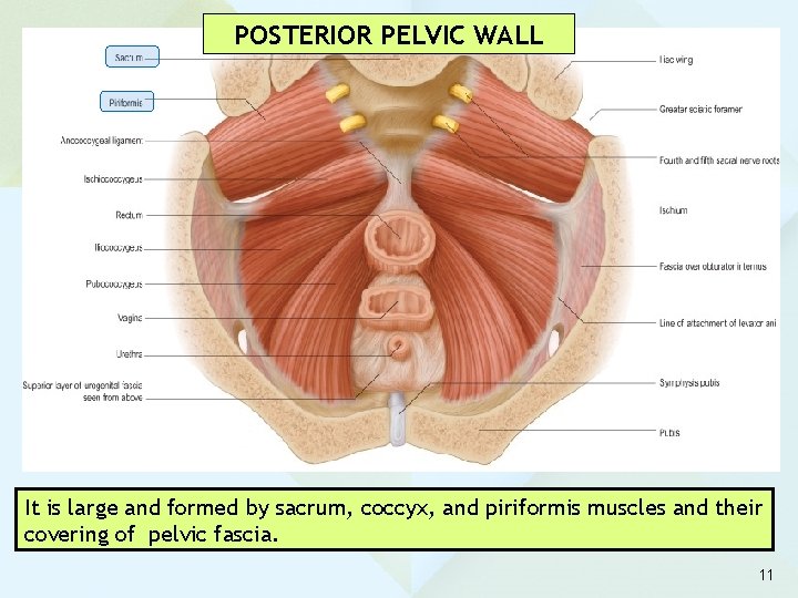
There are many muscles that form the pelvic floor, including puborectalis, pubococcygeus, iliococcygeus and coccygeus.
Its primary role is in arm movement,. • it consists of a sheet of fascia that lines the walls and floor of the pelvis. The fibers then decussate to meet with the fibers from the contralateral side, to form a sling around the distal parts of the pelvic organs. The pelvic cavity and perineum. The gluteus maximus attaches from the posterior iliac crest, posterolateral sacrum, and coccyx to the gluteal tuberosity and itb. Biceps of thigh large muscle enabling the leg to flex on the thigh and to rotate outwardly (outside the median axis) and the thigh to extend on the pelvis. The muscles of the pelvis form its floor. The gastrocnemius is the most superficial of the muscles and has two heads, medial and lateral. The levator ani muscles are the largest group of muscles in the pelvis. A diagram of the pelvis in medial view shows the muscles such as the psoas major, the iliac, piriformis and obturator internus muscles. There are two major groups of ligaments that provide nearly all the structure of the pelvis. • it covers the obturator internus, piriformis, levator ani and coccygeus muscles and is continuous with the transversalis fascia. Therefore, an appreciation of the female pelvic musculoskeletal anatomy is critical for understanding the pelvic support system.
• it covers the obturator internus, piriformis, levator ani and coccygeus muscles and is continuous with the transversalis fascia. A more detailed account of pelvic anatomy is best found in anatomy texts 1. The posterior abdominal wall is a musculoskeletal structure with numerous vascular and lymphatic structures formed by the lumbar vertebrae and their intervertebral discs, pelvic girdle, posterior abdominal wall muscles and their fascia. Most superficial, from anterior to posterior, are the adductor longus, gracilis, and adductor magnus. There are many muscles that form the pelvic floor, including puborectalis, pubococcygeus, iliococcygeus and coccygeus.
Hips Don T Lie Unique Hip Exercises For Power And Mobility Muscle Anatomy Hip Muscles Hip Workout from i.pinimg.com Pelvic anatomy is composed of two innominate (coxal) bones that articulate with the sacrum and proximal femora. The two largest muscles in this region include the gastrocnemius and the soleus. The posterior compartment is made up of a group of muscles called the hamstrings, including semitendinosus, semimembranosus and biceps femoris. Biceps of thigh large muscle enabling the leg to flex on the thigh and to rotate outwardly (outside the median axis) and the thigh to extend on the pelvis. Index 5 a distinction is made between the lesser or true pelvis inferior to the terminal line and the greater or false pelvis above it. A more detailed account of pelvic anatomy is best found in anatomy texts 1. The pelvic diaphragm is a wide but thin muscular layer of tissue that forms the inferior border of the abdominopelvic cavity. Sura) refers to the posterior portion of the lower leg.
Posterior hip musculature the posterior hip musculature comprises a group of muscles extending from the pelvic bone to the femur.
The pelvic floor is also known as the pelvic diaphragm. Index 5 a distinction is made between the lesser or true pelvis inferior to the terminal line and the greater or false pelvis above it. Spanning from the posterior pelvis to the proximal tibia and fibula, the posterior thigh muscles provide motion to both the femoroacetabular joint (hip joint) and tibiofemoral joint (knee joint). Pelvic anatomy is composed of two innominate (coxal) bones that articulate with the sacrum and proximal femora. The anterior fibers arise from the posterior surface of the pubic arch and travel posteriorly in the horizontal plane. The muscles of the pelvis form its floor. Arcus tendineus levator ani and the ischial spine These three muscles are collectively referred to as the hamstring muscles. The levator ani muscles consist of three. Its primary role is in arm movement,. Deep to these muscles are the adductor brevis anteriorly and the obturator externus posteriorly (fig. Therefore, an appreciation of the female pelvic musculoskeletal anatomy is critical for understanding the pelvic support system. There are many muscles that form the pelvic floor, including puborectalis, pubococcygeus, iliococcygeus and coccygeus.
The fibers then decussate to meet with the fibers from the contralateral side, to form a sling around the distal parts of the pelvic organs. Anatomy pelvis muscles muscles of the pelvis. Whenever someone talks about the pelvic floor in general, they are probably talking about these 5 muscles: Most superficial, from anterior to posterior, are the adductor longus, gracilis, and adductor magnus. The two largest muscles in this region include the gastrocnemius and the soleus.
Pelvic Floor Wikipedia from upload.wikimedia.org Biceps of thigh large muscle enabling the leg to flex on the thigh and to rotate outwardly (outside the median axis) and the thigh to extend on the pelvis. The pelvic diaphragm is a wide but thin muscular layer of tissue that forms the inferior border of the abdominopelvic cavity. Together with popliteus, flexor hallucis longus and flexor digitorum longus, it forms the deep group of muscles of the posterior compartment of leg. The pelvic cavity and perineum. The pubococcygeus is the intermediate part of the levator ani muscles. Spanning from the posterior pelvis to the proximal tibia and fibula, the posterior thigh muscles provide motion to both the femoroacetabular joint (hip joint) and tibiofemoral joint (knee joint). In the back of the torso, the latissimus dorsi is a large, rectangular muscle that extends from the lower back near the top of the pelvis to near the shoulder. Deep to these muscles are the adductor brevis anteriorly and the obturator externus posteriorly (fig.
These three muscles are collectively referred to as the hamstring muscles.
The gluteus maximus attaches from the posterior iliac crest, posterolateral sacrum, and coccyx to the gluteal tuberosity and itb. Deep to these muscles are the adductor brevis anteriorly and the obturator externus posteriorly (fig. The pelvic floor is also known as the pelvic diaphragm. The gastrocnemius is the most superficial of the muscles and has two heads, medial and lateral. Together with popliteus, flexor hallucis longus and flexor digitorum longus, it forms the deep group of muscles of the posterior compartment of leg. The pubococcygeus is the intermediate part of the levator ani muscles. Posterior hip musculature the posterior hip musculature comprises a group of muscles extending from the pelvic bone to the femur. Biceps of thigh large muscle enabling the leg to flex on the thigh and to rotate outwardly (outside the median axis) and the thigh to extend on the pelvis. The posterior abdominal wall is a musculoskeletal structure with numerous vascular and lymphatic structures formed by the lumbar vertebrae and their intervertebral discs, pelvic girdle, posterior abdominal wall muscles and their fascia. They have several functions, including helping to support the pelvic organs. A diagram of the pelvis in medial view shows the muscles such as the psoas major, the iliac, piriformis and obturator internus muscles. In the back of the torso, the latissimus dorsi is a large, rectangular muscle that extends from the lower back near the top of the pelvis to near the shoulder. The bony pelvis comes together to provide support for the pelvic muscles and connective tissues, which, in turn, provide attachments and support for the pelvic organs.
The pubococcygeus is the intermediate part of the levator ani muscles anatomy muscles pelvis. Arcus tendineus levator ani and the ischial spine